IMAGING BIOLOGICAL SURFACES
|
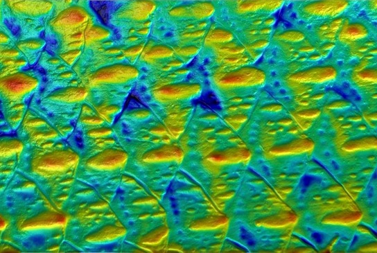
Surfaces provide many important functions when organisms interact with their surroundings, and I am interested in how evolution and function have shaped the diversity of organismal surfaces, and how we can accurately image biological surfaces to better understand their diversity. A major need in studying biological surfaces is finding a method that can deliver three-dimensional topographic information for surfaces that may be soft, wet, shiny, or transparent. To that end, I have pioneered the use of gel-based stereo profilometry to rapidly image biological surfaces in situ and in vivo. This method works on many biological surfaces but my research focuses on aquatic animals, especially fishes. At left are some images of scales from Osmerus mordax. See blog post on the 3D surface topography method I use here. |
EVOLUTIONARY PATTERNS OF FISH SURFACES
|
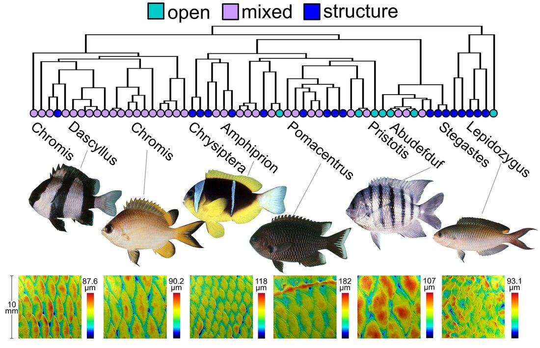
Biologists have long wondered why fish surfaces are so diverse. Fish surfaces come in a large variety of forms and although we think surfaces serve some particular functions (e.g. protection from predators, parasites, and disease), we do not understand how changes in surface form connect with changes in function and performance. I am working to explore this topic by using comparative studies of scale and surface morphology - combining modern phylogenetic comparative methods with 3D surface topography. So far, I've studied damselfish surfaces (a species-rich group of reef fishes) and shown that they respond to changes in ecology and body shape in ways that suggest ecological connections to scale morphology. Damselfishes also show differences in surface form when a species' flow ecology is different - suggesting a hydrodynamic role for scales. These results hold promise for continuing this line of research in more fish groups, and for generating new hypotheses for biomechanical testing. Understanding the generation and maintenance of diversity is a major question in evolutionary biology, and fish surfaces are an interesting system for exploring this topic.
|
|
BIOMECHANICS & HYDRODYNAMICS OF AQUATIC SURFACES
Fishes and other aquatic animals are constantly interacting with the viscous water around them that provides buoyancy and allows them to generate lift and drag. Learning about how aquatic animals move efficiently through water is a focus of both marine biology, comparative biomechanics, and bio-inspired engineering, and I have studied this topic throughout my work on biological surfaces in groups such as bony fishes, sharks, and cetaceans (whales and dolphins). In some cases I use topographic information to calculate if surfaces are likely to influence boundary-layer hydrodynamics, whereas in other cases I take a direct experimental approach to test how mucus causes drag reduction in fish skin. This area of research has implications for understanding form-function relationships for biological surfaces, but also for discovering new bio-inspired mechanisms for drag reduction that could be interest to engineering research and technology.

Boundary layer flow over fish scales.
|
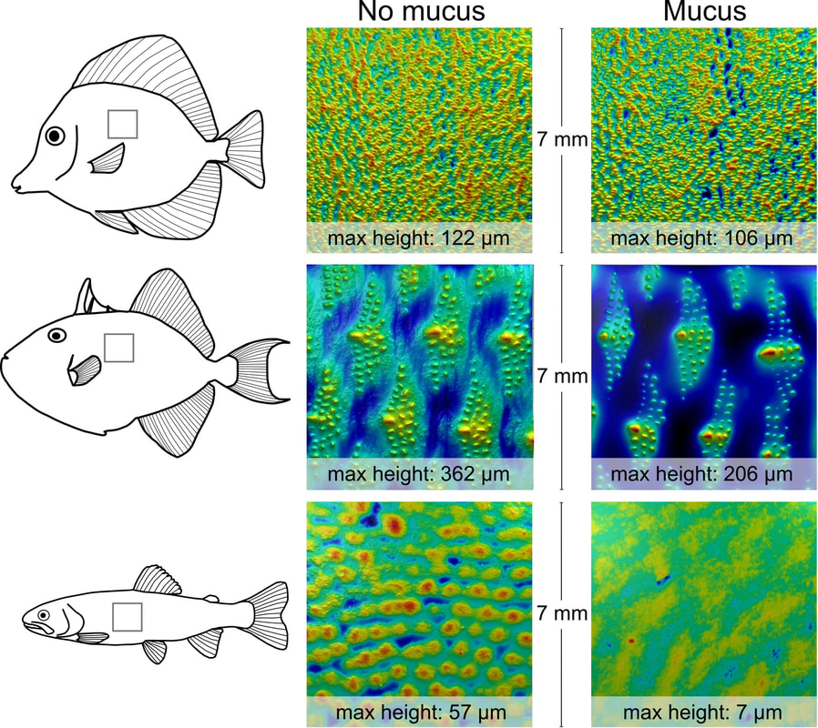
Surface of fishes with and without mucus; cool colors are low heights. Mucus has different effects, but often obscures the microstructure on fish scales.
|
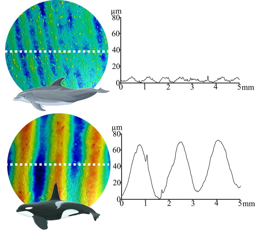
Ridges on cetacean skin are too small on most species to effect the boundary layer.
|
SCALE DIVERSITY IN FISHES
|
|
Scales are bony plates that cover the bodies of most fishes and many years of studies have shown us that scales show a myriad of forms in both different species and locations on the body. I have worked to modernize our understanding of scale diversity by using methods like micro computed tomography (µCT) scanning and gel-based profilometry to reveal undiscovered 3D diversity in fish scales. It is crucial to understand 3D structure of scales to allow us to better study the functional morphology of fish scales. My studies in this area have demonstrated that scales are diverse across different body regions according to a number of different morphological measurements. I also discovered that tunas show extreme diversity of scale shapes and sizes across their bodies, and interestingly, some tuna scales are thickened and filled with fat (shown in video at right) - possibly to act as surface insulation for the raised core temperatures of endothermic tunas.
|
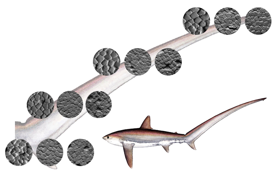
SHARK SKIN DIVERSITY AND HYDRODYNAMICS
|
Sharks have small complex scales called denticles that cover their bodies. Denticles have been of great interest to biologists and engineers alike in part because they are known to decrease drag and increase swimming performance under certain hydrodynamic conditions. My work in this area has mostly been with undergraduate collaborators and I have worked with them to study patterns of 3D structure across sharks bodies. We have discovered that shark denticles occur in many forms across the body but that they show repeated features from leading-edge to trailing-edge surfaces with respect to flow. These morphological patterns might indicate hydrodynamic tuning to local flows at these locations. I am also working to better connect denticle morphology and hydrodynamic function by directly measuring boundary layer flows (just a few millimeters from the skin) in live sharks.
|
|
FUNCTIONAL MORPHOLOGY OF TUNAS
Tuna lateral peduncle keels from µCT data.
|
Tunas are one of the most economically-important groups of fishes and their biology as large, high-performance, open-ocean, warm-bodied predators makes them biologically interesting and important as well. Tunas are thought of as highly specialized for swimming fast and they have a number of novel morphological features. I used µCT scanning to study some of these features and then created physical models of these features to test their hydrodynamic function using fish-like locomotion in a flow tunnel. Generally I found that tuna features like lateral peduncle keels and finlets decrease lateral forces and torques, which could make tuna swimming more efficient.
|
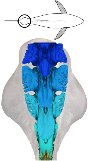
BIO-INSPIRED ADHESION
There are a handful of fish clades that have independently evolved the ability to adhere to underwater surfaces - sometimes to hitch a ride on another organism, or sometimes to simply stay-put in high-flow habitats. I have worked on a number of these systems to explore their adhesive performance and relevant morphology, and I have also collaborated with experts in bio-inspired robotics to both emulate and understand adhesive mechanisms and behavior. This work helps to build transferrable knowledge with potential uses in engineered technology. (Video below shows a volume rendering of part of the head and adhesive disc of a remora; from µCT data with soft-tissue contrast enhancement).
|
|
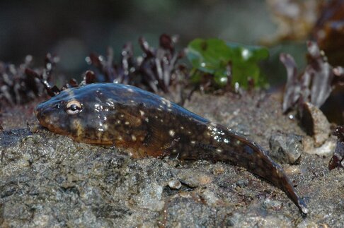
Northern clingfishes can adhere to rough and slippery surfaces using an adhesive disc on their belly. [Photo courtesy of Dr. Thomas Kleinteich]
|